HSCI researchers find that spinal cord repair relies on specialized cells

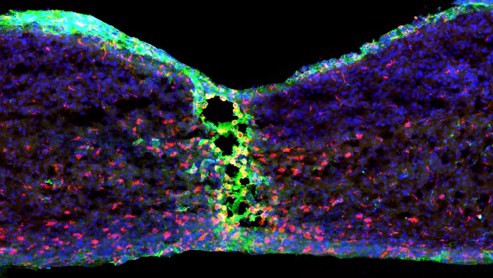
After spinal cord injury, the resulting scar tissue prevents nerve cells from reconnecting and hinders recovery. A new study by HSCI researchers at Boston Children’s Hospital has demonstrated a way to minimize scar formation in adult mice after a spinal cord injury. The research, published in the journal Nature, offers insights for new treatment approaches.
In adult mammals, axons do not regrow after spinal cord injury. Moreover, many types of harmful cells accumulate around the injury site, further interfering with the axons’ ability to reconnect and transmit signals between nerve cells. In this study, HSCI Principal Faculty member Zhigang He took a different approach to the problem, studying spinal cord injury and repair responses in 2-day-old mice.
“Unexpectedly, we found that the injury in these young pups lead to scar-free healing that permitted the growth of axons through the site of the lesion,” said He.
The researchers identified microglial cells — a type of immune cell found in the brain and spinal cord that remove damaged nerve cells and infection — as having an essential protective role in scar-free wound repair. When they studied a type of mouse that did not have normal numbers of microglia, they found that scar-free healing did not occur, and neither did axon regrowth.
The researchers found that microglia have at least two roles in scar-free healing. First, they help re-form bridges between the severed axon ends. Second, microglia from newborn mice — but not adult mice — produce a number of molecules that interfere with the action of certain harmful proteins.
“These protein inhibitors are involved in quickly tamping down the inflammatory response after spinal cord injury,” said He. “The microglia essentially orchestrated swift removal of harmful cell debris after injury and stopped inflammation.”
The researchers next tested whether axons could be regenerated in adult mice with spinal cord injuries. They found that transplanting microglia from newborn mice into adult mice with spinal cord lesions significantly improved healing and axon growth. They also saw similar results when transplanting adult microglia that were pre-treated with the helpful protein inhibitors.
“With these results, we are convinced that microglia are a primary organizer for this scar-free wound healing,” said He.
The researchers are now testing several types of protein inhibitors to see which may be most effective in boosting the scar-free healing potential of microglial cells.
This discovery may also help understanding of treatments for neurodegeneration in general, such as in Alzheimer’s disease, ALS, and Parkinson’s disease – conditions where microglia that are too active can have a negative impact.
“One of the key players in these diseases is microglia,” said He. His laboratory is now studying the role dysfunctional microglia may have in the ongoing destructive nerve damage seen in these diseases. “Perhaps we can find a gene that might convert those chronically active microglia to treat neurodegeneration.”
Read more
This story was originally published on the Boston Children’s Hospital website on October 8, 2020, under the title “Scar-free healing after spinal cord injury relies on specialized cells.”
Source article: Li, Y., He, X., Kawaguchi, R., et al. (2020). Microglia-organized scar-free spinal cord repair in neonatal mice. Nature. DOI: 10.1038/s41586-020-2795-6
This study was supported by the National Institutes of Health, a Ray W. Poppleton endowment, the Dr. Miriam and Sheldon G. Adelson Medical Research and the Craig H. Neilsen Foundation.
