 Harvard stem cell scientists have discovered that the clump of cells that gives rise to the embryonic heart also contains cells that form the heart’s plumbing, such as the aorta and the other great vessels.
While the discovery was made serendipitously by researchers working to illuminate the location of stem cells involved in embryonic heart formation in zebrafish, the finding, published in Nature Cell Biology, gives promise of explaining characteristics of congenital diseases in humans, such as DiGeorge syndrome, and may eventually lead to new therapeutic strategies.
The discovery was made by researchers, led by Caroline Burns, PhD, and Geoffrey Burns, PhD, both members of the Harvard Stem Cell Institute (HSCI) and Massachusetts General Hospital Cardiovascular Research Center, who genetically engineered zebrafish with glowing hearts to study early heart formation. The genetically modified fish were designed so that cells emitted yellow light if they expressed a gene associated with cardiac development, called nkx2.5.
“What we didn’t expect was to also see fluorescence within the pharyngeal arch arteries, which contribute to the formation of the great vessels of the heart,” said Caroline Burns. ”There’s virtually nothing in the literature about how these vessels are established in the first place, and what we found is that these arch arteries are coming from this region in the embryo that’s been classified as the heart field.”
Encouraging data gathered in collaboration with Richard Harvey, PhD, at the Victor Chang Research Institute in Australia, found that the arch artery cells found in zebrafish heart fields also lead to great vessel formation in mice, and likely other higher vertebrates. The researchers are looking to apply this new information to understand human disease, specifically in cases when connections between the arch arteries and the heart don’t properly form, causing health problems or embryonic death.
“There are examples of human patients with nkx2.5 mutations that have defective large vessels, so it’s possible that the reason theyhave these developmental defects is because of a genetic mutation,” said Geoffrey Burns. “We found that when you knockout the function of nkx2.5, you do not form these vessels, so it is required for the cells to form properly.”
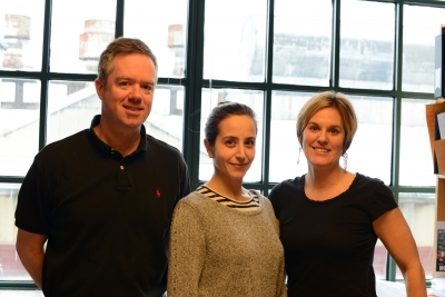
Burns Laboratory postdoctoral fellow Noëlle Paffett-Lugassy, PhD, the recipient of a 2011-2013 HSCI Training Grant, played a critical role in generating data for the study.
The work was also supported by a Harvard Stem Cell Institute Seed Grant and Young Investigator’s Award, the NIH National Heart, Lung and Blood Institute, the American Heart Association, the March of Dimes Foundation, the National Health and Medical Research Council of Australia, Atlantic Philanthropies, and the National Heart Foundation of Australia.
Citation: Heart field origin of great vessel precursors relies on nkx2.5-mediated vascuologenesis. Nature Cell Biology. Oct. 27, 2013